
Nur für die Forschung bestimmt
Sorafenib Tosylate (BAY 43-9006) Raf/VEGFR-Inhibitor
Kat.-Nr.: S1040
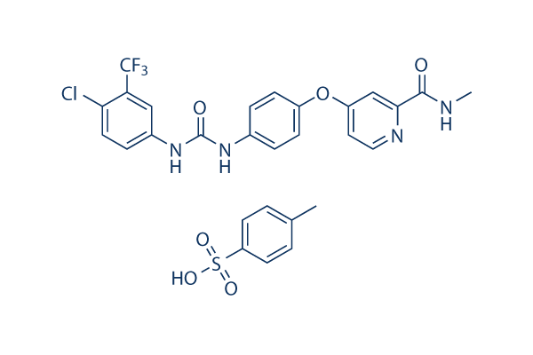
Chemische Struktur
Molekulargewicht: 637.03
Qualitätskontrolle (Quality Control)
Produkte, die oft zusammen verwendet werden mit Sorafenib Tosylate (BAY 43-9006)
| Verwandte Targets | ERK p38 MAPK JNK MEK Ras KRas S6 Kinase MAP4K TAK1 Mixed Lineage Kinase |
|---|---|
| Andere Raf Inhibitors | LY3009120 Exarafenib (KIN-2787) GDC-0879 Avutometinib (Ro5126766, CH5126766) PLX-4720 AZ 628 SB590885 TAK-632 GW5074 RAF265 (CHIR-265) |
Zellkultur, Behandlung & Arbeitskonzentration
(Cell Culture, Treatment & Working Concentration)
| Zelllinien | Assay-Typ | Konzentration | Inkubationszeit | Formulierung | Aktivitätsbeschreibung | PMID |
|---|---|---|---|---|---|---|
| MDA-MB-435 | Growth Inhibition Assay | 48 h | GI50=2 μM | 22560627 | ||
| UACC257 | Growth Inhibition Assay | 48 h | GI50=2 μM | 22560627 | ||
| MCF7 | Growth Inhibition Assay | 48 h | GI50=2.5 μM | 22560627 | ||
| EKVX | Growth Inhibition Assay | 48 h | GI50=2.5 μM | 22560627 | ||
| HT-29 | Growth Inhibition Assay | 48 h | GI50=2.5 μM | 22560627 | ||
| SNB19 | Growth Inhibition Assay | 48 h | GI50=3.2 μM | 22560627 | ||
| OVCAR3 | Growth Inhibition Assay | 48 h | GI50=3.2 μM | 22560627 | ||
| CAKI-1 | Growth Inhibition Assay | 48 h | GI50=3.2 μM | 22560627 | ||
| SW620 | Growth Inhibition Assay | 48 h | GI50=3.2 μM | 22560627 | ||
| TK10 | Growth Inhibition Assay | 48 h | GI50=5 μM | 22560627 | ||
| endothelial precursor cells | Function assay | Inhibition of endothelial cord area formation in endothelial precursor cells by CD31 cord area detection based phenotypic drug discovery based assay, IC50 = 0.00421 μM. | 22409666 | |||
| Sf9 | Function assay | Inhibition of GST-tagged recombinant human VEGFR2 expressed in Sf9 cells by radiometric assay, IC50 = 0.0125 μM. | 24368209 | |||
| endothelial precursor cells/ADSC cells | Toxicity assay | Toxicity in endothelial precursor cells co-cultured with stromal precursor ADSC cells by total nuclei count detection based phenotypic drug discovery based assay, EC50 = 5.46 μM. | 22409666 | |||
| endothelial precursor cells | Function assay | Inhibition of cell migration in endothelial precursor cells by Oris cell migration kit based phenotypic drug discovery based assay, IC50 = 16.7 μM. | 22409666 | |||
| RD | qHTS assay | qHTS of pediatric cancer cell lines to identify multiple opportunities for drug repurposing: Primary screen for RD cells | 29435139 | |||
| SK-N-SH | qHTS assay | qHTS of pediatric cancer cell lines to identify multiple opportunities for drug repurposing: Primary screen for SK-N-SH cells | 29435139 | |||
| OHS-50 | qHTS assay | qHTS of pediatric cancer cell lines to identify multiple opportunities for drug repurposing: Primary screen for OHS-50 cells | 29435139 | |||
| Klicken Sie hier, um weitere experimentelle Zellliniendaten anzuzeigen | ||||||
Chemische Informationen, Lagerung & Stabilität (Chemical Information, Storage & Stability)
| Molekulargewicht | 637.03 | Formel | C21H16ClF3N4O3.C7H8O3S |
Lagerung (Ab Erhaltdatum) | |
|---|---|---|---|---|---|
| CAS-Nr. | 475207-59-1 | SDF herunterladen | Lagerung von Stammlösungen |
|
|
| Synonyme | BAY 43-9006 tosylate,NSC-724772 tosylate | Smiles | CC1=CC=C(C=C1)S(=O)(=O)O.CNC(=O)C1=NC=CC(=C1)OC2=CC=C(C=C2)NC(=O)NC3=CC(=C(C=C3)Cl)C(F)(F)F | ||
Löslichkeit (Solubility)
|
In vitro |
DMSO
: 127 mg/mL
(199.36 mM)
Water : 0.01 mg/mL Ethanol : Insoluble |
Molaritätsrechner
|
In vivo |
|||||
In-vivo-Formulierungsrechner (Klare Lösung)
Schritt 1: Geben Sie unten die Informationen ein (Empfohlen: Ein zusätzliches Tier einplanen, um Verluste während des Experiments auszugleichen)
Schritt 2: Geben Sie die In-vivo-Formulierung ein (Dies ist nur der Rechner, nicht die Formulierung. Bitte kontaktieren Sie uns zuerst, wenn im Abschnitt Löslichkeit keine In-vivo-Formulierung angegeben ist.)
Berechnungsergebnisse:
Arbeitskonzentration: mg/ml;
Methode zur Herstellung der DMSO-Stammflüssigkeit: mg Wirkstoff voraufgelöst in μL DMSO ( Konzentration der Stammflüssigkeit mg/mL, Bitte kontaktieren Sie uns zuerst, wenn die Konzentration die DMSO-Löslichkeit der jeweiligen Wirkstoffcharge überschreitet. )
Methode zur Herstellung der In-vivo-Formulierung: Nehmen Sie μL DMSO Stammflüssigkeit, fügen Sie als Nächstes hinzuμL PEG300, mischen und aufklären, fügen Sie als Nächstes hinzuμL Tween 80, mischen und aufklären, fügen Sie als Nächstes hinzu μL ddH2O, mischen und aufklären.
Methode zur Herstellung der In-vivo-Formulierung: Nehmen Sie μL DMSO Stammflüssigkeit, fügen Sie als Nächstes hinzu μL Maisöl, mischen und aufklären.
Hinweis: 1. Bitte stellen Sie sicher, dass die Flüssigkeit klar ist, bevor Sie das nächste Lösungsmittel hinzufügen.
2. Achten Sie darauf, die Lösungsmittel in der richtigen Reihenfolge hinzuzufügen. Sie müssen sicherstellen, dass die im vorherigen Schritt erhaltene Lösung klar ist, bevor Sie das nächste Lösungsmittel hinzufügen. Physikalische Methoden wie Vortexen, Ultraschall oder ein warmes Wasserbad können zur Unterstützung des Lösungsvorgangs verwendet werden.
Wirkungsmechanismus (Mechanism of Action)
| Targets/IC50/Ki |
Raf-1
(Cell-free assay) 6 nM
VEGFR2/Flk1
(Cell-free assay) 15 nM
B-Raf
(Cell-free assay) 22 nM
B-Raf (V599E)
(Cell-free assay) 38 nM
PDGFRβ
(Cell-free assay) 57 nM
mPDGFRβ
(Cell-free assay) 57 nM
FLT3
(Cell-free assay) 58 nM
c-Kit
(Cell-free assay) 68 nM
VEGFR2
(Cell-free assay) 90 nM
FGFR1
(Cell-free assay) 580 nM
|
|---|---|
| In vitro |
Sorafenib tosylate inhibits both wild-type and V599E mutant B-Raf activity with IC50 of 22 nM and 38 nM, respectively. This compound also potently inhibits mVEGFR2 (Flk-1), mVEGFR3, mPDGFRβ, Flt3, and c-Kit with IC50 of 15 nM, 20 nM, 57 nM, 58 nM, and 68 nM, respectively. It weakly inhibits FGFR-1 with IC50 of 580 nM. This chemical is not active against ERK-1, MEK-1, EGFR, HER-2, IGFR-1, c-Met, PKB, PKA, cdk1/cyclinB, PKCα, PKCγ, and pim-1. It markedly inhibits VEGFR2 phosphorylation in NIH 3T3 cells with IC50 of 30 nM, and Flt-3 phosphorylation in HEK-293 cells with IC50 of 20 nM. It potently blocks MEK 1/2 and ERK 1/2 phosphorylation in most cell lines but not in A549 or H460 cells, while having no effect on inhibition of the PKB pathway. It inhibits the proliferation of HAoSMC and MDA-MB-231 cells with IC50 of 0.28 μM and 2.6 μM, respectively. In addition to inhibition of the RAF/MEK/ERK signaling pathway, it significantly inhibits the phosphorylation of eIF4E and down-regulates Mcl-1 levels in hepatocellular carcinoma (HCC) cells in a MEK/ERK-independent manner. It inhibits the proliferation of PLC/PRF/5 and HepG2 cells with IC50 of 6.3 μM and 4.5 μM, respectively, and leads to the significant induction of apoptosis. |
| Kinase-Assay |
Biochemische Assays
|
|
Rekombinante Baculoviren, die Raf-1 (Reste 305–648) und B-Raf (Reste 409–765) exprimieren, werden als Fusionsproteine gereinigt. Vollständiges humanes MEK-1 wird durch PCR erzeugt und als Fusionsprotein aus Escherichia coli-Lysaten gereinigt. Sorafenib-Tosylat wird zu einer Mischung aus Raf-1 (80 ng) oder B-Raf (80 ng) mit MEK-1 (1 μg) in Assay-Puffer [20 mM Tris (pH 8,2), 100 mM NaCl, 5 mM MgCl₂ und 0,15 % β-Mercaptoethanol] in einer Endkonzentration von 1 % DMSO. Der Raf-Kinase-Assay (Endvolumen 50 μl) wird durch Zugabe von 25 μl 10 μM γ[33P]ATP (400 Ci/mol) gestartet und 25 Minuten lang bei 32 °C inkubiert. Phosphoryliertes MEK-1 wird durch Filtration auf einer Phosphocellulosematte gesammelt, und 1 %ige Phosphorsäure wird verwendet, um ungebundene Radioaktivität wegzuwaschen. Nach dem Trocknen durch Mikrowellenerwärmung wird ein β-Plattenzähler verwendet, um die filtergebundene Radioaktivität zu quantifizieren. Die Kinasedomäne des humanen VEGFR2 (KDR) wird aus Sf9-Lysaten exprimiert und gereinigt. Zeitaufgelöste Fluoreszenzenergietransfer-Assays für VEGFR2 werden in 96-Well-Platten im Format des zeitaufgelösten Fluoreszenzenergietransfers durchgeführt. Die endgültigen Reaktionsbedingungen sind wie folgt: 1 bis 10 μM ATP, 25 nM Poly-GT-Biotin, 2 nM europiummarkierter Phospho-(p)-Tyr-Antikörper (PY20), 10 nM APC, 1 bis 7 nM zytoplasmatische Kinasedomäne in Endkonzentrationen von 1 % DMSO, 50 mM HEPES (pH 7,5), 10 mmol/L MgCl₂, 0,1 mmol/L EDTA, 0,015 % Brij-35, 0,1 mg/ml BSA und 0,1 % β-Mercaptoethanol. Das Reaktionsvolumen beträgt 100 μl und die Reaktion wird durch Zugabe des Enzyms gestartet. Die Platten werden sowohl bei 615 als auch bei 665 nm auf einem Perkin-Elmer VictorV Multilabel-Zähler etwa 1,5 bis 2,0 Stunden nach Reaktionsbeginn ausgelesen. Das Signal wird als Verhältnis berechnet: (665 nm/615 nm) × 10.000 für jede Vertiefung. Zur Ermittlung des IC50-Werts wird diese Verbindung vor der Enzymaktivierung hinzugefügt. Eine 50-fache Stammlösung wird mit dieser Chemikalie hergestellt, die in einer 50 %igen DMSO/50 %igen destillierten Wasserlösung 1:3 seriell verdünnt wird. Die Endkonzentrationen dieser Verbindung liegen zwischen 10 μM und 4,56 nM in 1 % DMSO.
|
|
| In vivo |
Oral administration of Sorafenib tosylate (~60 mg/kg) demonstrates broad spectrum, dose-dependent anti-tumor activity against a variety of human tumor xenograft models including MDA-MB-231, Colo-205, HT-29, DLD-1, NCI-H460, and A549, with no evidence of toxicity. In association with the anti-tumor efficacy, this compound treatment potently inhibits MEK 1/2 phosphorylation and pERK 1/2 levels in HT-29 and MDA-MB-231 xenografts but not in Colo-205 xenografts, and significantly suppresses tumor microvessel area (MVA) and microvessel density (MVD) in MDA MB-231, HT-29 and Colo-205 tumor xenografts. This compound treatment produces dose-dependent growth inhibition of PLC/PRF/5 tumor xenografts in SCID mice with TGIs of 49% and 78% at 10 mg/kg and 30 mg/kg, respectively, consistent with the inhibition of ERK and eIF4E phosphorylation, reduction of the microvessel area, and induction of tumor cell apoptosis. |
Referenzen |
|
Anwendungen (Applications)
| Methoden | Biomarker | Bilder | PMID |
|---|---|---|---|
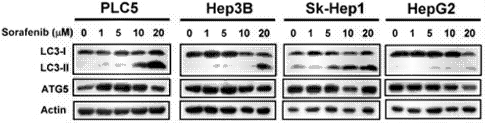
| Western blot | LC3-I / LC-3II / ATG5 p-STAT3 / STAT3/ Mcl-1 β-catenin / Survivin / Mcl-1 / PTMA pERK / ERK p-PKM2(Y105) / PMK2 / Caspase-9 RET(pY1016) / VEGFR2(pY1214) / MEK1(pT292) / ERK(pY204) Cyclin D1 |
|
23392173 |
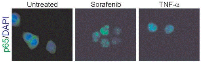
| Immunofluorescence | p65 cytochrome c |
|
22286758 |
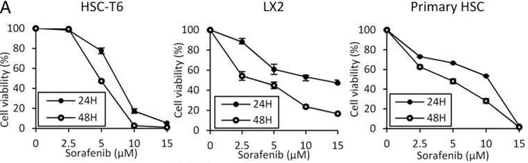
| Growth inhibition assay | Cell viability |
|
26039995 |
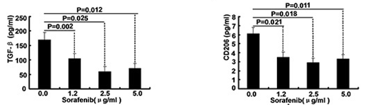
| ELISA | TGF-beta / CD206 Caspase-9 / Caspase-3 |
|
26158762 |
Informationen zu klinischen Studien (Clinical Trial Information)
(Daten von https://clinicaltrials.gov, aktualisiert am 2026-08-26)
| NCT-Nummer | Rekrutierung | Bedingungen | Sponsor/Kollaboratoren | Startdatum | Phases |
|---|---|---|---|---|---|
| NCT07787429 | RECRUITING | Osteosarcoma |
Anna Raciborska |
2026-04-01 | PHASE2 |
| NCT05468359 | RECRUITING | Refractory Solid Tumor; Hepatocellular Carcinoma; Malignant Solid Tumor; Pediatric Cancer; Pediatric Solid Tumor; Fibrolamellar Carcinoma |
St. Jude Children's Research Hospital |
2022-11-07 | PHASE1; PHASE2 |
| NCT02559778 | RECRUITING | Neuroblastoma |
Giselle Sholler |
2015-09 | PHASE2 |
| NCT06972641 | RECRUITING | AML; MDS |
Ruijin Hospital |
2025-06-10 | PHASE2; PHASE3 |
| NCT07560566 | RECRUITING | Hepato Cellular Carcinoma (HCC); Hand Foot Skin Syndrome |
National Taiwan University Hospital |
2025-12-09 | PHASE2 |
| NCT06227221 | NOT_YET_RECRUITING | Type 1 Diabetes |
Second Xiangya Hospital of Central South University |
2026-02 | PHASE2 |

Die Produkte sind nur für Forschungszwecke bestimmt. Nicht zur Anwendung am Menschen. Kein Verkauf an Patienten.
©Copyright 2013 Selleck Chemicals. Alle Rechte vorbehalten.