
Nur für die Forschung bestimmt
Dovitinib (TKI-258) FLT3 Inhibitor
Kat.-Nr.: S1018
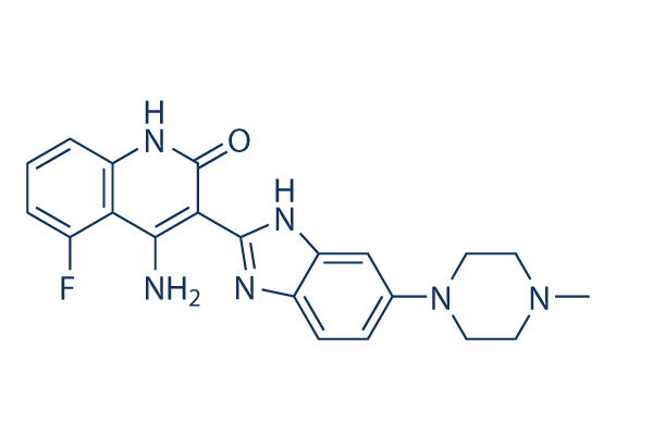
Chemische Struktur
Molekulargewicht: 392.43
Qualitätskontrolle (Quality Control)
| Verwandte Targets | EGFR VEGFR JAK FGFR PDGFR Src HIF HER2 FLT Bcr-Abl |
|---|---|
| Andere FLT3 Inhibitors | UNC2025 Crenolanib (CP-868596) Dovitinib (TKI258) Lactate monohydrate Tandutinib (MLN518) ENMD-2076 KW-2449 AST-487 (NVP-AST487) TCS 359 G-749 FF-10101 |
Zellkultur, Behandlung & Arbeitskonzentration
(Cell Culture, Treatment & Working Concentration)
| Zelllinien | Assay-Typ | Konzentration | Inkubationszeit | Formulierung | Aktivitätsbeschreibung | PMID |
|---|---|---|---|---|---|---|
| SupB15 | Growth Inhibition Assay | IC50=0.449 μM | 25202073 | |||
| SupB15-R | Growth Inhibition Assay | IC50=0.558 μM | 25202073 | |||
| BaF3-pSRα | Growth Inhibition Assay | IC50=0.668 μM | 25202073 | |||
| BaF3-p210Bcr-Abl | Growth Inhibition Assay | IC50=0.692 μM | 25202073 | |||
| BaF3-p210Bcr-Abl-T315I | Growth Inhibition Assay | IC50=2.626 μM | 25202073 | |||
| CCRF-CEM | Growth Inhibition Assay | IC50=0.398 μM | 25202072 | |||
| CEM/C2 | Growth Inhibition Assay | IC50=1.125 μM | 25202072 | |||
| Nalm-6 | Growth Inhibition Assay | IC50=0.382 μM | 25202072 | |||
| SEM-K2 | Growth Inhibition Assay | IC50=0.022 μM | 25202072 | |||
| HB-1119 | Growth Inhibition Assay | IC50=0.028 μM | 25202072 | |||
| RS4:11 | Growth Inhibition Assay | IC50=2.81 μM | 25202072 | |||
| Nalm-6 | Apoptosis Assay | 2 μM | 24/48 h | induces apoptosis resulting in about 72% of cell death after 24 h treatment and 81% after 48 h | 25202072 | |
| SEM-K2 | Apoptosis Assay | 0.1/1 μM | 24 h | induces early apoptosis of SEM-K2 cells at 0.1 μM after 24 h | 25202072 | |
| HCT-116 | Growth Inhibition Assay | IC50=3.050.58 μM | 24495750 | |||
| HT-29 | Growth Inhibition Assay | IC50=5.21.93 μM | 24495750 | |||
| SW-480 | Growth Inhibition Assay | IC50=4.330.47 μM | 24495750 | |||
| CaCO2 | Growth Inhibition Assay | IC50=3.230.64 μM | 24495750 | |||
| LS174T | Growth Inhibition Assay | IC50=4.330.47 μM | 24495750 | |||
| HEC-1A | Function Assay | 0.05/0.1/0.5 μM | 72 h | causes a decrease in STAT3, ERK, and AKT phosphorylation | 24495750 | |
| AN3CA | Function Assay | 0.05/0.1/0.5 μM | 72 h | causes a decrease in STAT3, ERK, and AKT phosphorylation | 24495750 | |
| MFE-296 | Function Assay | 0.05/0.1/0.5 μM | 72 h | causes a decrease in STAT3, ERK, and AKT phosphorylation | 24495750 | |
| UMC3 | Cell Viability Assay | 1-10 μM | 72 h | inhibits cell growth in a dose dependent manner | 24325461 | |
| 5637 | Cell Viability Assay | 1-10 μM | 72 h | inhibits cell growth in a dose dependent manner | 24325461 | |
| HU456 | Cell Viability Assay | 1-10 μM | 72 h | inhibits cell growth in a dose dependent manner | 24325461 | |
| MGHU4 | Cell Viability Assay | 1-10 μM | 72 h | inhibits cell growth in a dose dependent manner | 24325461 | |
| HT1376 | Cell Viability Assay | 1-10 μM | 72 h | inhibits cell growth in a dose dependent manner | 24325461 | |
| RT112 | Cell Viability Assay | 1-10 μM | 72 h | inhibits cell growth in a dose dependent manner | 24325461 | |
| T24 | Cell Viability Assay | 1-10 μM | 72 h | inhibits cell growth in a dose dependent manner | 24325461 | |
| BFTC905 | Cell Viability Assay | 1-10 μM | 72 h | inhibits cell growth in a dose dependent manner | 24325461 | |
| TCC-SUP | Cell Viability Assay | 1-10 μM | 72 h | inhibits cell growth in a dose dependent manner | 24325461 | |
| RT4 | Cell Viability Assay | 1-10 μM | 72 h | inhibits cell growth in a dose dependent manner | 24325461 | |
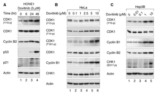
| HONE1 | Growth Inhibition Assay | 0.1-10 μM | 48 h | induces G2/M delay in a concentration-dependent manner | 24238094 | |
| HNE1 | Growth Inhibition Assay | 0.1-10 μM | 48 h | induces G2/M delay in a concentration-dependent manner | 24238094 | |
| CNE2 | Growth Inhibition Assay | 0.1-10 μM | 48 h | induces G2/M delay in a concentration-dependent manner | 24238094 | |
| C666-1 | Growth Inhibition Assay | 0.1-10 μM | 48 h | induces G2/M delay in a concentration-dependent manner | 24238094 | |
| HeLa | Growth Inhibition Assay | 0.1-10 μM | 24 h | induces G2/M arrest in a concentration-dependent manner | 24238094 | |
| Hep3B | Growth Inhibition Assay | 0.1-10 μM | 24 h | induces G2 arrest | 24238094 | |
| HepG2 | Growth Inhibition Assay | 48 h | IC50=2.727 ± 0.429 μM | 23546591 | ||
| Hep3B | Growth Inhibition Assay | 48 h | IC50=4.223 ± 0.839 μM | 23546591 | ||
| PLC/PRF5 | Growth Inhibition Assay | 48 h | IC50=16.120 ± 4.001 μM | 23546591 | ||
| Huh7 | Growth Inhibition Assay | 48 h | IC50=15.007 ± 7.334 μM | 23546591 | ||
| HepG2 | Growth Inhibition Assay | 72 h | IC50=1.200 ± 0.226 μM | 23546591 | ||
| Hep3B | Growth Inhibition Assay | 72 h | IC50=0.892 ± 0.044 μM | 23546591 | ||
| PLC/PRF5 | Growth Inhibition Assay | 72 h | IC50=3.110 ± 0.337 μM | 23546591 | ||
| Huh7 | Growth Inhibition Assay | 72 h | IC50=3.980 ± 0.803 μM | 23546591 | ||
| MFE280 | Growth Inhibition Assay | IC50=0.42 ± 0.06 μM | 23443805 | |||
| AN3CA | Growth Inhibition Assay | IC50=0.50 ± 0.10 μM | 23443805 | |||
| HEC155 | Growth Inhibition Assay | IC50=0.66 ± 0.09 μM | 23443805 | |||
| MFE296 | Growth Inhibition Assay | IC50=0.66 ± 0.19 μM | 23443805 | |||
| SPAC1S | Growth Inhibition Assay | IC50=0.77 ± 0.08 μM | 23443805 | |||
| RL952 | Growth Inhibition Assay | IC50=0.93 ± 0.01 μM | 23443805 | |||
| EN1 | Growth Inhibition Assay | IC50=1.02 ± 0.25 μM | 23443805 | |||
| SNGII | Growth Inhibition Assay | IC50=1.24 ± 0.28 μM | 23443805 | |||
| ISHIKAWA | Growth Inhibition Assay | IC50=1.30 ± 0.11 μM | 23443805 | |||
| HEC1A | Growth Inhibition Assay | IC50=1.34 ± 0.30 μM | 23443805 | |||
| KLE | Growth Inhibition Assay | IC50=1.37 ± 0.02 μM | 23443805 | |||
| SNGM | Growth Inhibition Assay | IC50=1.42 ± 0.13 μM | 23443805 | |||
| USPC2 | Growth Inhibition Assay | IC50=1.62 ± 0.01 μM | 23443805 | |||
| EN | Growth Inhibition Assay | IC50=1.66 ± 0.01 μM | 23443805 | |||
| MFE319 | Growth Inhibition Assay | IC50=1.87 ± 0.45 μM | 23443805 | |||
| EFE184 | Growth Inhibition Assay | IC50=2.04 ± 0.13 μM | 23443805 | |||
| ECC1 | Growth Inhibition Assay | IC50=2.07 ± 0.01 μM | 23443805 | |||
| HEC1B | Growth Inhibition Assay | IC50=2.57 ± 0.23 μM | 23443805 | |||
| USPC1 | Growth Inhibition Assay | IC50=2.60 ± 0.13 μM | 23443805 | |||
| SPAC1L | Growth Inhibition Assay | IC50=3.06 ± 1.14 μM | 23443805 | |||
| HUVEC | Cell Viability Assay | 0-25 μM | 72 h | DMSO | inhibits cell growth in a dose dependent manner | 23228017 |
| HMVEC | Cell Viability Assay | 0-25 μM | 72 h | DMSO | inhibits cell growth in a dose dependent manner | 23228017 |
| MHCC-97H | Cell Viability Assay | 0-25 μM | 72 h | DMSO | inhibits cell growth in a dose dependent manner | 23228017 |
| SMMC7721 | Cell Viability Assay | 0-25 μM | 72 h | DMSO | inhibits cell growth in a dose dependent manner | 23228017 |
| Huh-7 | Apoptosis Assay | 0-12.5 μM | 24 h | DMSO | sensitizes HCC cells to TRAIL- and tigatuzumab-induced apoptosis in a dose-dependent manner | 22230479 |
| Sk-Hep1 | Apoptosis Assay | 0-12.5 μM | 24 h | DMSO | sensitizes HCC cells to TRAIL- and tigatuzumab-induced apoptosis in a dose-dependent manner | 22230479 |
| Hep3B | Apoptosis Assay | 0-12.5 μM | 24 h | DMSO | sensitizes HCC cells to TRAIL- and tigatuzumab-induced apoptosis in a dose-dependent manner | 22230479 |
| PLC5 | Apoptosis Assay | 0-12.5 μM | 24 h | DMSO | sensitizes HCC cells to TRAIL- and tigatuzumab-induced apoptosis in a dose-dependent manner | 22230479 |
| PLC5 | Cell Viability Assay | 0-15 μM | 72 h | reduces cell viability in a dose-dependent manner | 22180308 | |
| Hep3B | Cell Viability Assay | 0-15 μM | 72 h | reduces cell viability in a dose-dependent manner | 22180308 | |
| Sk-Hep1 | Cell Viability Assay | 0-15 μM | 72 h | reduces cell viability in a dose-dependent manner | 22180308 | |
| Huh-7 | Cell Viability Assay | 0-15 μM | 72 h | reduces cell viability in a dose-dependent manner | 22180308 | |
| PLC5 | Apoptosis Assay | 0-15 μM | 24 h | increases apoptotic cell death in a dose-dependent manner | 22180308 | |
| Hep3B | Apoptosis Assay | 0-15 μM | 24 h | increases apoptotic cell death in a dose-dependent manner | 22180308 | |
| Sk-Hep1 | Apoptosis Assay | 0-15 μM | 24 h | increases apoptotic cell death in a dose-dependent manner | 22180308 | |
| Huh-7 | Apoptosis Assay | 0-15 μM | 24 h | increases apoptotic cell death in a dose-dependent manner | 22180308 | |
| PLC5 | Function Assay | 0-10 μM | 24 h | causes dose-dependent DNA fragmentation | 22180308 | |
| Hep3B | Function Assay | 0-10 μM | 24 h | causes dose-dependent DNA fragmentation | 22180308 | |
| Sk-Hep1 | Function Assay | 0-10 μM | 24 h | causes dose-dependent DNA fragmentation | 22180308 | |
| Huh-7 | Function Assay | 0-10 μM | 24 h | causes dose-dependent DNA fragmentation | 22180308 | |
| SW780 | Growth Inhibition Assay | 5 d | IC50=50 nM | 21119661 | ||
| RT112 | Growth Inhibition Assay | 5 d | IC50=15 nM | 21119661 | ||
| RT4 | Growth Inhibition Assay | 5 d | IC50=5 nM | 21119661 | ||
| JMSU1 | Growth Inhibition Assay | 5 d | IC50=50 nM | 21119661 | ||
| J82 | Growth Inhibition Assay | 5 d | IC50=1400 nM | 21119661 | ||
| 97-7 | Growth Inhibition Assay | 5 d | IC50=1000 nM | 21119661 | ||
| RT112 | Function Assay | 500 nM | 24 h | increases the proportion of cells in G1 accompanied by a decrease in S and G2/M phases | 21119661 | |
| RT4 | Function Assay | 500 nM | 24 h | increases the proportion of cells in G1 accompanied by a decrease in S and G2/M phases | 21119661 | |
| MGH-U3 | Function Assay | 500 nM | 24 h | increases the proportion of cells in G1 accompanied by a decrease in S and G2/M phases | 21119661 | |
| SW780 | Function Assay | 500 nM | 24 h | increases the proportion of cells in G1 accompanied by a decrease in S and G2/M phases | 21119661 | |
| 97-7 | Function Assay | 500 nM | 24 h | increases the proportion of cells in G1 accompanied by a decrease in S and G2/M phases | 21119661 | |
| J807C | Cell Viability Assay | 0-400 nM | 48 h | inhibits cell growth in a dose dependent manner | 15598814 | |
| Y373C | Cell Viability Assay | 0-400 nM | 48 h | inhibits cell growth in a dose dependent manner | 15598814 | |
| K650E | Cell Viability Assay | 0-400 nM | 48 h | inhibits cell growth in a dose dependent manner | 15598814 | |
| G384D | Cell Viability Assay | 0-400 nM | 48 h | inhibits cell growth in a dose dependent manner | 15598814 | |
| F384L | Cell Viability Assay | 0-400 nM | 48 h | inhibits cell growth in a dose dependent manner | 15598814 | |
| KMS11 | Growth Inhibition Assay | 72 h | IC50=90 nM | 15598814 | ||
| KMS18 | Growth Inhibition Assay | 72 h | IC50=550 nM | 15598814 | ||
| OPM2 | Growth Inhibition Assay | 72 h | IC50=90 nM | 15598814 | ||
| H929 | Growth Inhibition Assay | 72 h | IC50> 2500 nM | 15598814 | ||
| 8226 | Growth Inhibition Assay | 72 h | IC50> 2500 nM | 15598814 | ||
| U266 | Growth Inhibition Assay | 72 h | IC50> 2500 nM | 15598814 | ||
| KM12L4A | Function assay | Inhibition of VEGFR2 phosphorylation expressed in human KM12L4A cells by Western blot analysis, EC50=0.046μM | 19113866 | |||
| KM12L4A | Function assay | Inhibition of PDGFRbeta phosphorylation expressed in human KM12L4A cells Western blot analysis, EC50=0.051μM | 19113866 | |||
| KM12L4A | Function assay | Inhibition of FGFR1 phosphorylation expressed in human KM12L4A cells by Western blot analysis, EC50=0.166μM | 19113866 | |||
| insect cells | Function assay | 1 to 4 hrs | Inhibition of recombinant PDGFRbeta (unknown origin) expressed in baculovirus infected insect cells using biotinylated peptide substrate GGLFDDPSYVNVQNL in presence of ATP incubated for 1 to 4 hrs by time resolved fluorescence assay, IC50=0.001μM | 27914362 | ||
| Sf9 | Function assay | 1 to 4 hrs | Inhibition of recombinant human N-terminal GST/His6-tagged c-KIT (544 to 976 residues) expressed in baculovirus infected sf9 cells using biotinylated peptide substrate GGLFDDPSYVNVQNL in presence of ATP incubated for 1 to 4 hrs by time resolved fluorescen, IC50=0.001μM | 27914362 | ||
| Sf9 | Function assay | 1 to 4 hrs | Inhibition of recombinant human N-terminal GST/His6-tagged FLT3 (571 to 993 residues) expressed in baculovirus infected sf9 cells using biotinylated peptide substrate GGLFDDPSYVNVQNL in presence of ATP incubated for 1 to 4 hrs by time resolved fluorescenc, IC50=0.001μM | 27914362 | ||
| insect cells | Function assay | 1 to 4 hrs | Inhibition of recombinant FGFR1 (unknown origin) expressed in baculovirus infected insect cells using biotinylated peptide substrate GGGGQDGKDYIVLPI in presence of ATP incubated for 1 to 4 hrs by time resolved fluorescence assay, IC50=0.008μM | 27914362 | ||
| insect cells | Function assay | 1 to 4 hrs | Inhibition of recombinant VEGFR3 (unknown origin) expressed in baculovirus infected insect cells using biotinylated peptide substrate GGGGQDGKDYIVLPI in presence of ATP incubated for 1 to 4 hrs by time resolved fluorescence assay, IC50=0.008μM | 27914362 | ||
| insect cells | Function assay | 1 to 4 hrs | Inhibition of recombinant VEGFR1 (unknown origin) expressed in baculovirus infected insect cells using biotinylated peptide substrate GGGGQDGKDYIVLPI in presence of ATP incubated for 1 to 4 hrs by time resolved fluorescence assay, IC50=0.01μM | 27914362 | ||
| insect cells | Function assay | 1 to 4 hrs | Inhibition of recombinant VEGFR2 (unknown origin) expressed in baculovirus infected insect cells using biotinylated peptide substrate GGGGQDGKDYIVLPI in presence of ATP incubated for 1 to 4 hrs by time resolved fluorescence assay, IC50=0.013μM | 27914362 | ||
| TC32 | qHTS assay | qHTS of pediatric cancer cell lines to identify multiple opportunities for drug repurposing: Primary screen for TC32 cells | 29435139 | |||
| SJ-GBM2 | qHTS assay | qHTS of pediatric cancer cell lines to identify multiple opportunities for drug repurposing: Primary screen for SJ-GBM2 cells | 29435139 | |||
| A673 | qHTS assay | qHTS of pediatric cancer cell lines to identify multiple opportunities for drug repurposing: Primary screen for A673 cells | 29435139 | |||
| SK-N-MC | qHTS assay | qHTS of pediatric cancer cell lines to identify multiple opportunities for drug repurposing: Primary screen for SK-N-MC cells | 29435139 | |||
| BT-37 | qHTS assay | qHTS of pediatric cancer cell lines to identify multiple opportunities for drug repurposing: Primary screen for BT-37 cells | 29435139 | |||
| NB-EBc1 | qHTS assay | qHTS of pediatric cancer cell lines to identify multiple opportunities for drug repurposing: Primary screen for NB-EBc1 cells | 29435139 | |||
| U-2 OS | qHTS assay | qHTS of pediatric cancer cell lines to identify multiple opportunities for drug repurposing: Primary screen for U-2 OS cells | 29435139 | |||
| Saos-2 | qHTS assay | qHTS of pediatric cancer cell lines to identify multiple opportunities for drug repurposing: Primary screen for Saos-2 cells | 29435139 | |||
| SK-N-SH | qHTS assay | qHTS of pediatric cancer cell lines to identify multiple opportunities for drug repurposing: Primary screen for SK-N-SH cells | 29435139 | |||
| NB1643 | qHTS assay | qHTS of pediatric cancer cell lines to identify multiple opportunities for drug repurposing: Primary screen for NB1643 cells | 29435139 | |||
| LAN-5 | qHTS assay | qHTS of pediatric cancer cell lines to identify multiple opportunities for drug repurposing: Primary screen for LAN-5 cells | 29435139 | |||
| BT-12 | qHTS assay | qHTS of pediatric cancer cell lines to identify multiple opportunities for drug repurposing: Primary screen for BT-12 cells | 29435139 | |||
| Rh18 | qHTS assay | qHTS of pediatric cancer cell lines to identify multiple opportunities for drug repurposing: Primary screen for Rh18 cells | 29435139 | |||
| OHS-50 | qHTS assay | qHTS of pediatric cancer cell lines to identify multiple opportunities for drug repurposing: Primary screen for OHS-50 cells | 29435139 | |||
| RD | qHTS assay | qHTS of pediatric cancer cell lines to identify multiple opportunities for drug repurposing: Primary screen for RD cells | 29435139 | |||
| insect cells | Function assay | 1 to 4 hrs | Inhibition of recombinant FGFR1 (unknown origin) expressed in baculovirus infected insect cells using GGGGQDGKDYIVLPI as substrate after 1 to 4 hrs by time-resolved fluorescence assay, IC50=0.008μM | 30503938 | ||
| NCI-H1703 | Function assay | 10 uM | 24 hrs | Inhibition of TNIK in human NCI-H1703 cells transfected with lentiviral vector 7TFP assessed as reduction of GSK3 inhibitor X activated TNIK-mediated Wnt/TCF/beta-catenin-dependent transcription at 10 uM after 24 hrs by luciferase reporter assay | ChEMBL | |
| LoVo | Cytotoxicity assay | 10 uM | 72 hrs | Cytotoxicity against Wnt/beta-catenin signalling dependent human LoVo cells assessed as cell viability at 10 uM after 72 hrs by ATPlite assay | ChEMBL | |
| HCT116 | Cytotoxicity assay | 10 uM | 72 hrs | Cytotoxicity against Wnt/beta-catenin signalling dependent human HCT116 cells assessed as cell viability at 10 uM after 72 hrs by ATPlite assay | ChEMBL | |
| Klicken Sie hier, um weitere experimentelle Zellliniendaten anzuzeigen | ||||||
Chemische Informationen, Lagerung & Stabilität (Chemical Information, Storage & Stability)
| Molekulargewicht | 392.43 | Formel | C21H21FN6O |
Lagerung (Ab Erhaltdatum) | |
|---|---|---|---|---|---|
| CAS-Nr. | 405169-16-6 | SDF herunterladen | Lagerung von Stammlösungen |
|
|
| Synonyme | CHIR-258 | Smiles | CN1CCN(CC1)C2=CC3=C(C=C2)N=C(N3)C4=C(C5=C(C=CC=C5F)NC4=O)N | ||
Löslichkeit (Solubility)
|
In vitro |
DMSO
: 30 mg/mL
(76.44 mM)
Water : Insoluble Ethanol : Insoluble |
Molaritätsrechner
|
In vivo |
|||||
In-vivo-Formulierungsrechner (Klare Lösung)
Schritt 1: Geben Sie unten die Informationen ein (Empfohlen: Ein zusätzliches Tier einplanen, um Verluste während des Experiments auszugleichen)
Schritt 2: Geben Sie die In-vivo-Formulierung ein (Dies ist nur der Rechner, nicht die Formulierung. Bitte kontaktieren Sie uns zuerst, wenn im Abschnitt Löslichkeit keine In-vivo-Formulierung angegeben ist.)
Berechnungsergebnisse:
Arbeitskonzentration: mg/ml;
Methode zur Herstellung der DMSO-Stammflüssigkeit: mg Wirkstoff voraufgelöst in μL DMSO ( Konzentration der Stammflüssigkeit mg/mL, Bitte kontaktieren Sie uns zuerst, wenn die Konzentration die DMSO-Löslichkeit der jeweiligen Wirkstoffcharge überschreitet. )
Methode zur Herstellung der In-vivo-Formulierung: Nehmen Sie μL DMSO Stammflüssigkeit, fügen Sie als Nächstes hinzuμL PEG300, mischen und aufklären, fügen Sie als Nächstes hinzuμL Tween 80, mischen und aufklären, fügen Sie als Nächstes hinzu μL ddH2O, mischen und aufklären.
Methode zur Herstellung der In-vivo-Formulierung: Nehmen Sie μL DMSO Stammflüssigkeit, fügen Sie als Nächstes hinzu μL Maisöl, mischen und aufklären.
Hinweis: 1. Bitte stellen Sie sicher, dass die Flüssigkeit klar ist, bevor Sie das nächste Lösungsmittel hinzufügen.
2. Achten Sie darauf, die Lösungsmittel in der richtigen Reihenfolge hinzuzufügen. Sie müssen sicherstellen, dass die im vorherigen Schritt erhaltene Lösung klar ist, bevor Sie das nächste Lösungsmittel hinzufügen. Physikalische Methoden wie Vortexen, Ultraschall oder ein warmes Wasserbad können zur Unterstützung des Lösungsvorgangs verwendet werden.
Wirkungsmechanismus (Mechanism of Action)
| Targets/IC50/Ki |
FLT3
(Cell-free assay) 1 nM
c-Kit
(Cell-free assay) 2 nM
FGFR1
(Cell-free assay) 8 nM
VEGFR3/FLT4
(Cell-free assay) 8 nM
FGFR3
(Cell-free assay) 9 nM
VEGFR1/FLT1
(Cell-free assay) 10 nM
VEGFR2/Flk1
(Cell-free assay) 13 nM
PDGFRβ
(Cell-free assay) 27 nM
CSF-1R/c-Fms
(Cell-free assay) 36 nM
|
|---|---|
| In vitro |
Dovitinib (TKI-258) potently inhibits the FGF-stimulated growth of WT and F384L-FGFR3-expressing B9 cells with IC50 of 25 nM. In addition, it inhibits proliferation of B9 cells expressing each of the various activated mutants of FGFR3. Interestingly, there are minimal observed differences in the sensitivity of the different FGFR3 mutations to this compound, with the IC50 ranging from 70 to 90 nM for each of the various mutations. IL-6-dependent B9 cells containing vector only (B9-MINV cells are resistant to its inhibitory activity at concentrations up to 1 μM. It inhibits cell proliferation of KMS11 (FGFR3-Y373C), OPM2 (FGFR3-K650E), and KMS18 (FGFR3-G384D) cells with IC50 of 90 nM (KMS11 and OPM2) and 550 nM, respectively. The compound inhibits FGF-mediated ERK1/2 phosphorylation and induces cytotoxicity in FGFR3-expressing primary MM cells. BMSCs does confer a modest degree of resistance with 44.6% growth inhibition for cells treated with 500 nM Dovitinib and cultured on stroma compared with 71.6% growth inhibition for cells grown without BMSCs. It inhibits proliferation of M-NFS-60, an M-CSF growth-driven mouse myeloblastic cell line with a median effective concentration (EC50) of 220 nM. Treatment of SK-HEP1 cells with this compound results in a dose-dependent reduction in cell number and G2/M phase arrest with reduction in the G0/G1 and S phases, inhibition of anchorage-independent growth and blockage of bFGF-induced cell motility. The IC50 for it in SK-HEP1 cells is approximately 1.7 μM. It also significantly reduces the basal phosphorylation levels of FGFR-1, FGFR substrate 2α (FRS2-α) and ERK1/2 but not Akt in both SK-HEP1 and 21-0208 cells. In 21-0208 HCC cells, it significantly inhibits bFGF-induced phosphorylation of FGFR-1, FRS2-α, ERK1/2 but not Akt. |
| Kinase-Assay |
In-vitro-Kinase-Assays
|
|
Die 50%igen Hemmkonzentrationen (IC50-Werte) für die Hemmung von RTKs durch Dovitinib (TKI-258) werden in einem zeitaufgelösten Fluoreszenz- (TRF) oder radioaktiven Format bestimmt, indem die Hemmung der Phosphatübertragung auf ein Substrat durch das jeweilige Enzym gemessen wird. Die Kinase-Domänen von FGFR3, FGFR1, PDGFRβ und VEGFR1-3 werden in 50 mM HEPES (N-2-Hydroxyethylpiperazin-N′-2-ethansulfonsäure), pH 7,0, 2 mM MgCl2, 10 mM MnCl2, 1 mM NaF, 1 mM Dithiothreitol (DTT), 1 mg/mL Rinderserumalbumin (BSA), 0,25 μM biotinyliertem Peptidsubstrat (GGGGQDGKDYIVLPI) und 1 bis 30 μM Adenosintriphosphat (ATP) in Abhängigkeit vom Km für das jeweilige Enzym untersucht. Die ATP-Konzentrationen liegen bei oder knapp unter Km. Für c-KIT- und FLT3-Reaktionen wird der pH-Wert auf 7,5 erhöht, mit 0,2 bis 8 μM ATP in Gegenwart von 0,25 bis 1 μM biotinyliertem Peptidsubstrat (GGLFDDPSYVNVQNL). Die Reaktionen werden bei Raumtemperatur 1 bis 4 Stunden inkubiert und das phosphorylierte Peptid auf Streptavidin-beschichteten Mikrotiterplatten, die Stoppreaktionspuffer (25 mM EDTA [Ethylendiamintetraessigsäure], 50 mM HEPES, pH 7,5) enthalten, eingefangen. Phosphoryliertes Peptid wird mit dem DELFIA TRF-System unter Verwendung eines Europium-markierten Anti-Phosphotyrosin-Antikörpers PT66 gemessen. Die Konzentration dieser Verbindung für IC50 wird mittels nichtlinearer Regression mit der XL-Fit-Datenanalysesoftware Version 4.1 (IDBS) berechnet. Die Hemmung der Kinaseaktivität des Kolonie-stimulierenden Faktor-1-Rezeptors (CSF-1R), PDGFRα, Insulinrezeptors (InsR) und Insulin-ähnlichen Wachstumsfaktorrezeptors 1 (IGFR1) wird bei ATP-Konzentrationen nahe dem Km für ATP bestimmt.
|
|
| In vivo |
Dovitinib (TKI-258) induces both cytostatic and cytotoxic responses in vivo resulting in regression of FGFR3-expressing tumors. It shows a dose- and exposure-dependent inhibition of target receptor tyrosine kinases (RTKs) expressed in tumor xenografts. This compound potently inhibits tumor growth of six HCC lines. Inhibition of angiogenesis correlated with inactivation of FGFR/PDGFRβ/VEGFR2 signaling pathways. In an orthotopic model, it potently inhibits primary tumor growth and lung metastasis and significantly prolonged mouse survival. Administration of Dovitinib results in significant tumor growth inhibition and tumor regressions, including large, established tumors (500-1,000 mm3). |
Referenzen |
|
Anwendungen (Applications)
| Methoden | Biomarker | Bilder | PMID |
|---|---|---|---|
| Western blot | CDK1 / p-CDK1 / p53 / p21 p-PDGFR-β / PDGFR-β / p-ERK / ERK p-VEGFR-2 / VEGFR-2 / p-FGFR-1 / FGFR-1 p-STAT3 / STAT3 / Mcl-1 / LC3 / Beclin 1 / p62 |
|
24238094 |
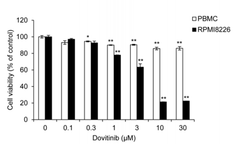
| Growth inhibition assay | Cell viability |
|
28467797 |
Informationen zu klinischen Studien (Clinical Trial Information)
(Daten von https://clinicaltrials.gov, aktualisiert am 2026-06-01)
| NCT-Nummer | Rekrutierung | Bedingungen | Sponsor/Kollaboratoren | Startdatum | Phases |
|---|---|---|---|---|---|
| NCT05571969 | TERMINATED | Advanced Solid Tumors |
Allarity Therapeutics |
2023-03-08 | PHASE1 |
| NCT05571969 | Recruiting | Advanced Solid Tumors |
Allarity Therapeutics|Amarex Clinical Research |
2023-02-20 | Phase 1 |
| NCT00831792 | COMPLETED | Prostate Cancer |
M.D. Anderson Cancer Center |
2010-04-07 | PHASE2 |
| NCT01994590 | TERMINATED | Prostate Cancer |
M.D. Anderson Cancer Center |
2014-05-19 | PHASE2 |
| NCT01732107 | TERMINATED | Bladder Cancer |
Noah Hahn, M.D. |
2013-03 | PHASE2 |
| NCT02116803 | COMPLETED | Solid Tumors |
Novartis Pharmaceuticals |
2014-05-28 | PHASE2; PHASE3 |

Die Produkte sind nur für Forschungszwecke bestimmt. Nicht zur Anwendung am Menschen. Kein Verkauf an Patienten.
©Copyright 2013 Selleck Chemicals. Alle Rechte vorbehalten.