
Nur für die Forschung bestimmt
BIIB021 HSP Inhibitor
Kat.-Nr.: S1175
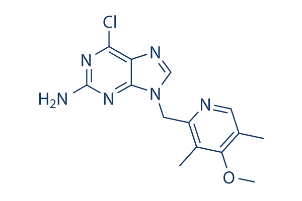
Chemische Struktur
Molekulargewicht: 318.76
Qualitätskontrolle (Quality Control)
Zellkultur, Behandlung & Arbeitskonzentration
(Cell Culture, Treatment & Working Concentration)
| Zelllinien | Assay-Typ | Konzentration | Inkubationszeit | Formulierung | Aktivitätsbeschreibung | PMID |
|---|---|---|---|---|---|---|
| MCF7 | Function assay | Inhibition of HSP90-mediated client protein HER2 degradation in human MCF7 cells, IC50=0.038μM | 20055425 | |||
| BT474 | Function assay | Binding affinity to Hsp90 nucleotide binding domain in human BT474 cells, IC50=0.14μM | 20608738 | |||
| MCF7 | Function assay | Inhibition of HSP90alpha in human MCF7 cells assessed as degradation of Her-2, EC50=0.038μM | 22938030 | |||
| NCI-H295 | Cytotoxicity assay | 120 mg/kg | 5 days | Cytotoxicity against human NCI-H295 cells overexpressing PGP xenografted in athymic mouse assessed as inhibition of tumor growth at 120 mg/kg, po qd for 5 days per week for 4 weeks | 22938030 | |
| Sf9 | Function assay | 3 hrs | Binding affinity to human N-terminal polyHis-tagged HSP90alpha (D9 to E236) alpha-helix conformation expressed in insect sf9 cells after 3 hrs by fluorescence polarization assay, Ki=0.002μM | 24332488 | ||
| Sf9 | Function assay | 3 hrs | Binding affinity to human N-terminal polyHis-tagged HSP90beta (D9 to E236) expressed in insect sf9 cells after 3 hrs by fluorescence polarization assay, Ki=0.004μM | 24332488 | ||
| NCI-H1299 | Function assay | 12 hrs | Reduction in oxygen consumption rate in human NCI-H1299 cells incubated for 12 hrs | 25383915 | ||
| HeLa | Function assay | 10 uM | 6 hrs | Inhibition of HSP90 in human HeLa cells assessed as induction of chk1 degradation at 10 uM after 6 hrs by Western blot method | 28816449 | |
| HeLa | Function assay | 10 uM | 6 hrs | Inhibition of HSP90 in human HeLa cells assessed as induction of Akt degradation at 10 uM after 6 hrs by Western blot method | 28816449 | |
| HeLa | Function assay | 10 uM | 6 hrs | Inhibition of HSP90 in human HeLa cells assessed as induction of HSP70 protein expression at 10 uM after 6 hrs by Western blot method | 28816449 | |
| PC3 | Function assay | 10 uM | 6 hrs | Inhibition of HSP90 in human PC3 cells assessed as induction of chk1 degradation at 10 uM after 6 hrs by Western blot method | 28816449 | |
| PC3 | Function assay | 10 uM | 6 hrs | Inhibition of HSP90 in human PC3 cells assessed as induction of Akt degradation at 10 uM after 6 hrs by Western blot method | 28816449 | |
| PC3 | Function assay | 10 uM | 6 hrs | Inhibition of HSP90 in human PC3 cells assessed as induction of HSP70 protein expression at 10 uM after 6 hrs by Western blot method | 28816449 | |
| TC32 | qHTS assay | qHTS of pediatric cancer cell lines to identify multiple opportunities for drug repurposing: Primary screen for TC32 cells | 29435139 | |||
| SJ-GBM2 | qHTS assay | qHTS of pediatric cancer cell lines to identify multiple opportunities for drug repurposing: Primary screen for SJ-GBM2 cells | 29435139 | |||
| A673 | qHTS assay | qHTS of pediatric cancer cell lines to identify multiple opportunities for drug repurposing: Primary screen for A673 cells | 29435139 | |||
| SK-N-MC | qHTS assay | qHTS of pediatric cancer cell lines to identify multiple opportunities for drug repurposing: Primary screen for SK-N-MC cells | 29435139 | |||
| NB-EBc1 | qHTS assay | qHTS of pediatric cancer cell lines to identify multiple opportunities for drug repurposing: Primary screen for NB-EBc1 cells | 29435139 | |||
| SK-N-SH | qHTS assay | qHTS of pediatric cancer cell lines to identify multiple opportunities for drug repurposing: Primary screen for SK-N-SH cells | 29435139 | |||
| NB1643 | qHTS assay | qHTS of pediatric cancer cell lines to identify multiple opportunities for drug repurposing: Primary screen for NB1643 cells | 29435139 | |||
| LAN-5 | qHTS assay | qHTS of pediatric cancer cell lines to identify multiple opportunities for drug repurposing: Primary screen for LAN-5 cells | 29435139 | |||
| Rh18 | qHTS assay | qHTS of pediatric cancer cell lines to identify multiple opportunities for drug repurposing: Primary screen for Rh18 cells | 29435139 | |||
| OHS-50 | qHTS assay | qHTS of pediatric cancer cell lines to identify multiple opportunities for drug repurposing: Primary screen for OHS-50 cells | 29435139 | |||
| Rh41 | qHTS assay | qHTS of pediatric cancer cell lines to identify multiple opportunities for drug repurposing: Primary screen for Rh41 cells | 29435139 | |||
| HCT116 | Antiproliferative assay | 48 hrs | Antiproliferative activity against human HCT116 cells after 48 hrs by sulforhodamine B assay, GI50=0.15μM | 29567459 | ||
| A549 | Antiproliferative assay | 48 hrs | Antiproliferative activity against human A549 cells after 48 hrs by sulforhodamine B assay, GI50=0.33μM | 29567459 | ||
| NCI-H1975 | Antiproliferative assay | 48 hrs | Antiproliferative activity against human NCI-H1975 cells after 48 hrs by sulforhodamine B assay, GI50=0.38μM | 29567459 | ||
| HL60 | Antiproliferative assay | 48 hrs | Antiproliferative activity against human HL60 cells after 48 hrs by sulforhodamine B assay, GI50=0.59μM | 29567459 | ||
| HL60 | Function assay | 1 uM | 24 hrs | Inhibition of HSP90 in human HL60 cells assessed as induction of HSP70 expression at 1 uM after 24 hrs by Western blot analysis | 29567459 | |
| HL60 | Function assay | 1 uM | 24 hrs | Inhibition of HSP90 in human HL60 cells assessed as downregulation of phosphorylated Akt expression at 1 uM after 24 hrs by Western blot analysis | 29567459 | |
| HL60 | Function assay | 1 uM | 24 hrs | Inhibition of HSP90 in human HL60 cells assessed as downregulation of phosphorylated STAT3 expression at 1 uM after 24 hrs by Western blot analysis | 29567459 | |
| HL60 | Function assay | 1 uM | 24 hrs | Inhibition of HDAC in human HL60 cells assessed as upregulation of acetyl-alpha-tubulin levels at 1 uM after 24 hrs by Western blot analysis | 29567459 | |
| HL60 | Function assay | 1 uM | 24 hrs | Inhibition of HDAC in human HL60 cells assessed as upregulation of acetylated histone H3 levels at 1 uM after 24 hrs by Western blot analysis | 29567459 | |
| MCF7 | Antiproliferative assay | Antiproliferative activity against human MCF7 cells, IC50=0.31μM | 31663736 | |||
| Klicken Sie hier, um weitere experimentelle Zellliniendaten anzuzeigen | ||||||
Chemische Informationen, Lagerung & Stabilität (Chemical Information, Storage & Stability)
| Molekulargewicht | 318.76 | Formel | C14H15ClN6O |
Lagerung (Ab Erhaltdatum) | |
|---|---|---|---|---|---|
| CAS-Nr. | 848695-25-0 | SDF herunterladen | Lagerung von Stammlösungen |
|
|
| Synonyme | CNF2024 | Smiles | CC1=CN=C(C(=C1OC)C)CN2C=NC3=C2N=C(N=C3Cl)N | ||
Löslichkeit (Solubility)
|
In vitro |
DMSO
: 64 mg/mL
(200.77 mM)
Ethanol : 12 mg/mL Water : Insoluble |
Molaritätsrechner
|
In vivo |
|||||
In-vivo-Formulierungsrechner (Klare Lösung)
Schritt 1: Geben Sie unten die Informationen ein (Empfohlen: Ein zusätzliches Tier einplanen, um Verluste während des Experiments auszugleichen)
Schritt 2: Geben Sie die In-vivo-Formulierung ein (Dies ist nur der Rechner, nicht die Formulierung. Bitte kontaktieren Sie uns zuerst, wenn im Abschnitt Löslichkeit keine In-vivo-Formulierung angegeben ist.)
Berechnungsergebnisse:
Arbeitskonzentration: mg/ml;
Methode zur Herstellung der DMSO-Stammflüssigkeit: mg Wirkstoff voraufgelöst in μL DMSO ( Konzentration der Stammflüssigkeit mg/mL, Bitte kontaktieren Sie uns zuerst, wenn die Konzentration die DMSO-Löslichkeit der jeweiligen Wirkstoffcharge überschreitet. )
Methode zur Herstellung der In-vivo-Formulierung: Nehmen Sie μL DMSO Stammflüssigkeit, fügen Sie als Nächstes hinzuμL PEG300, mischen und aufklären, fügen Sie als Nächstes hinzuμL Tween 80, mischen und aufklären, fügen Sie als Nächstes hinzu μL ddH2O, mischen und aufklären.
Methode zur Herstellung der In-vivo-Formulierung: Nehmen Sie μL DMSO Stammflüssigkeit, fügen Sie als Nächstes hinzu μL Maisöl, mischen und aufklären.
Hinweis: 1. Bitte stellen Sie sicher, dass die Flüssigkeit klar ist, bevor Sie das nächste Lösungsmittel hinzufügen.
2. Achten Sie darauf, die Lösungsmittel in der richtigen Reihenfolge hinzuzufügen. Sie müssen sicherstellen, dass die im vorherigen Schritt erhaltene Lösung klar ist, bevor Sie das nächste Lösungsmittel hinzufügen. Physikalische Methoden wie Vortexen, Ultraschall oder ein warmes Wasserbad können zur Unterstützung des Lösungsvorgangs verwendet werden.
Wirkungsmechanismus (Mechanism of Action)
| Targets/IC50/Ki |
HSP90
(Cell-free assay) 1.7 nM(Ki)
|
|---|---|
| In vitro |
BIIB021 binds in the ATP-binding pocket of Hsp90, interferes with Hsp90 chaperone function, and results in client protein degradation and tumor growth inhibition. This compound inhibits tumor cell (BT474, MCF-7, N87, HT29, H1650, H1299, H69 and H82) proliferation with IC50 from 0.06-0.31 μM. It induces the degradation of Hsp90 client proteins including HER-2, Akt, and Raf-1 and up-regulated expression of the heat shock proteins Hsp70 and Hsp27. This chemical inhibits Hodgkin's lymphoma cells (KM-H2, L428, L540, L540cy, L591, L1236 and DEV) with IC50 from 0.24-0.8 μM. It shows low activity in lymphocytes from healthy individuals. This compound inhibits the constitutive activity of NF-κB despite defective IκB. It induces the expression of ligands for the activating NK cell receptor NKG2D on Hodgkin's lymphoma cells resulting in an increased susceptibility to NK cell–mediated killing. This chemical enhanced the in vitro radiosensitivity of HNSCCA cell lines (UM11B and JHU12) with a corresponding reduction in the expression of key radioresponsive proteins, increased apoptotic cells and enhance G2 arrest. It is considerably more active than 17-AAG against adrenocortical carcinoma H295R, both in vitro and in vivo. The cytotoxic activity of this compound is not influenced by loss of NQO1 or Bcl-2 overexpression, molecular lesions that do not prevent client loss but are nonetheless associated with reduced cell killing by 17-AAG. It is also active in 17-AAG resistant cell lines (NIH-H69, MES SA Dx5, NCI-ADR-RES, Nalm6 and etc.).
|
| Kinase-Assay |
Hsp90-Bindungsassay
|
|
Für Fluoreszenzpolarisation-Kompetitionsmessungen wird die FITC-Geldanamycin-Sonde (20 nM) 3 Stunden lang bei Raumtemperatur mit 2 mM TCEP reduziert, wonach die Lösung aliquotiert und bei -80 °C gelagert wird, bis sie verwendet wird. Rekombinantes humanes Hsp90α (0,8 nM) und reduziertes FITC-Geldanamycin (2 nM) werden in einer 96-Well-Mikrotiterplatte 3 Stunden lang bei Raumtemperatur in Gegenwart eines Assay-Puffers inkubiert, der 20 mM HEPES (pH 7,4), 50 mM KCl, 5 mM MgCl2, 20 mM Na2MoO4, 2 mM DTT, 0,1 mg/mL BGG und 0,1 % (v/v) CHAPS enthält. Nach dieser Vorinkubation wird diese Verbindung in 100 % DMSO in Endkonzentrationen von 0,2 nM bis 10 μM (Endvolumen 100 μL, 2 % DMSO) hinzugefügt. Die Reaktion wird 16 Stunden lang bei Raumtemperatur inkubiert und die Fluoreszenz wird dann in einem Analyst-Plattenleser bei einer Anregung von 485 nm und einer Emission von 535 nm gemessen. Hohe und niedrige Kontrollen enthielten kein diese Chemikalie bzw. kein Hsp90. Die Daten werden an eine Vier-Parameter-Kurve angepasst und der IC50-Wert wird generiert.
|
|
| In vivo |
Oral administration of BIIB021 leads to tumor growth inhibition in many tumor xenograft models including N87, BT474, CWR22, U87, SKOV3 and Panc-1. This compound effectively inhibits growth of L540cy tumor at a dose of 120 mg/kg. It significantly enhances antitumor growth effect of radiation in JHU12 xenograft.
|
Referenzen |
|
Informationen zu klinischen Studien (Clinical Trial Information)
(Daten von https://clinicaltrials.gov, aktualisiert am 2015-10-02)
| NCT-Nummer | Rekrutierung | Bedingungen | Sponsor/Kollaboratoren | Startdatum | Phases |
|---|---|---|---|---|---|
| NCT01004081 | COMPLETED | Breast Cancer |
Biogen |
2009-11 | PHASE2 |
| NCT01017198 | COMPLETED | Advanced Solid Tumors |
Biogen |
2009-11 | PHASE1 |
| NCT00618735 | COMPLETED | Advanced Solid Tumors |
Biogen |
2008-02 | PHASE1 |
| NCT00618319 | COMPLETED | GIST |
Biogen |
2008-02 | PHASE2 |
| NCT01017198 | Completed | Advanced Solid Tumors |
Biogen |
2009-11 | Phase 1 |
| NCT01004081 | Completed | Breast Cancer |
Biogen |
2009-11 | Phase 2 |

Die Produkte sind nur für Forschungszwecke bestimmt. Nicht zur Anwendung am Menschen. Kein Verkauf an Patienten.
©Copyright 2013 Selleck Chemicals. Alle Rechte vorbehalten.